What it involves
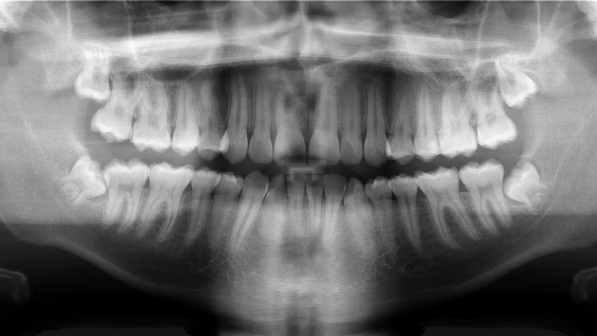
Dental X-rays are images of the teeth, roots and surrounding bone. Common types include bitewing images, which show the crowns of upper and lower back teeth, periapical images of whole teeth, and panoramic images of the whole jaw. They help a dentist see decay between teeth, infection, gum disease and the position of unerupted teeth.
Who it is for
They are used when the dentist needs information that cannot be seen by looking in the mouth, such as before treatment, when investigating pain or to monitor a problem. The dentist chooses X-rays based on your needs and history rather than at fixed intervals for everyone.
How long it usually takes
The exposure itself takes seconds, and the appointment time is usually a few minutes for a single image. A full panoramic image takes slightly longer.
What to expect
You will be asked about pregnancy and wear a protective apron or other protection if the practice uses one. You bite on a small holder or stand still while a machine moves around your head. The dentist then explains what the images show.
Aftercare
There is no aftercare. X-rays involve a small dose of ionising radiation, and dentists aim to use the lowest dose that answers the clinical question. Tell the dentist if you have had recent X-rays elsewhere so they can avoid repeating them where possible.
When to seek it
A dentist may recommend X-rays if you have toothache, a new problem, are starting treatment or have a history of decay or gum disease. You can ask why an image is needed.
What affects the cost
Cost depends on the type and number of images, whether digital equipment is used and whether they are part of a larger examination. HealthNaro shows a price only where a clinic has supplied a figure with a source and date.
This page is general information and not medical advice. In an emergency, contact your local emergency number.
Sources: World Health Organization; NHS; NHS.